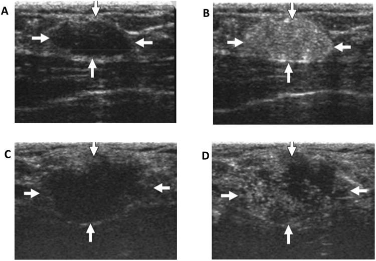
Figure 2.

(A) Unenhanced ultrasound image shows homogenously hypoechoic oval shaped solid mass (arrows) in fibroglandular tissue of breast with adenoma (22 years old women); (B) Contrast enhanced ultrasound image of same area; (C) Unenhanced ultrasound image shows lobulated hypoechoic mass (arrows) in fibroglandular tissue of papillotubular carcinoma (38 years old woman); (D) Contrast enhanced ultrasound image of same area shows clear internal defects. (Reprinted from Ref 32. Copyright 2014 Miyamoto, Y. et al – permission pending).
