
Figure 6.
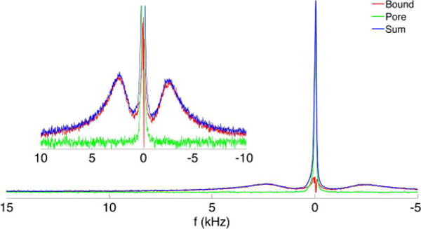
2H spectra showing the bound and pore D2O components (inset is magnified vertically and truncated). Pore water (narrow central peak in green) is calculated by subtracting the bound water spectrum obtained by inversion-recovery nulling of pore water (the split peaks shown in red) from the fully relaxed spectrum (shown in blue). This spectrum is taken from a specimen from a 27 year old female donor with the osteonal axis orthogonal to B0. Splitting of 4.8 kHz is observed, consistent with the orientation-dependent splitting observed by Ong et al. (9).