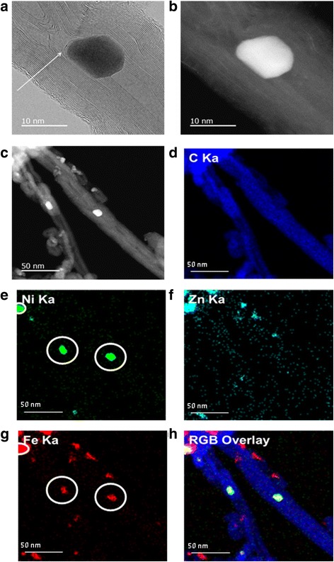
Fig. 4.

a STEM bright field image of single ZnFe2O4/mCNT. Arrow indicates the internal walls of mCNT partly coating ZnFe2O4 particle. b STEM dark field image of single ZnFe2O4/mCNT. c STEM annular dark-field image of ZnFe2O4/mCNTs. d The EDS x-ray maps of e nickel, the circles indicate Ni is agglomerated in the presence of ZnFe2O4. f Zinc. g Iron, the circles indicate the presence of iron in the same locations of Ni. h Display of the three specified maps as red, green, and blue overlays—representing iron, nickel, and carbon phase distributions
