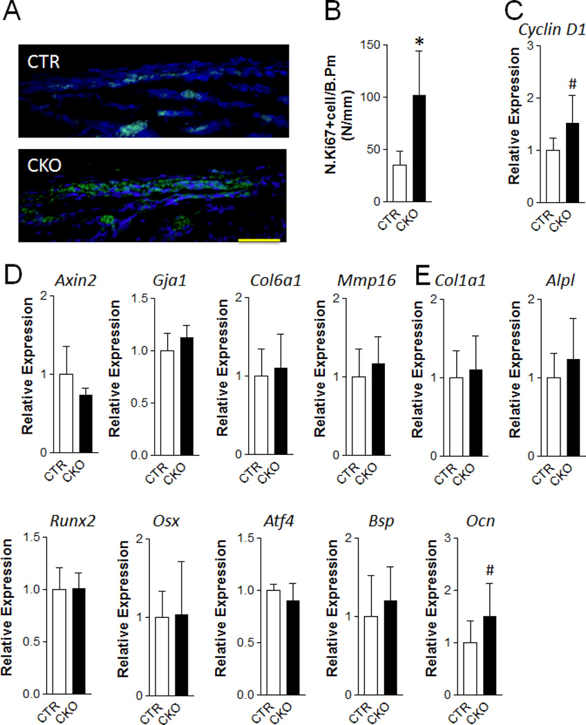
Figure 5. TSC1 deletion leads to increased osteoprogenitor cell proliferation at early postnatal stage.
(A, B) Immunofluorescence staining with anti-Ki67 antibody was performed in the frontal bone of one-week-old CTR and CKO mice. (A) Representative fluorescent images in the extracranial periosteum area. Scale bar=50µm. (B) Ki67 positive cell number per bone perimeter. *p<0.05, n=5 per group. (C–E) Quantitative-PCR analysis of the mRNA expression of cell cycle G1/S transition gene Cyclin D1 (C), Wnt signaling target genes: Axin2, Gja143, Col6a1, Mmp16 (D), and osteoblast differentiation markers: Col1a1, Alpl, Runx2, Osx, Atf4, Bsp, Ocn (E) of frontal bone of one-week-old CTR and CKO mice. #p<0.001, n=11–12 per group. Data were presented as mean ± SD.

