Figure 8.
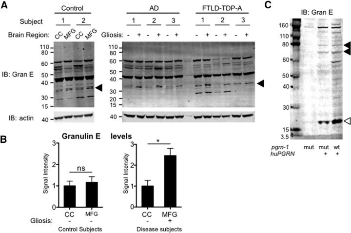
A granulin cleavage product is over-represented in diseased brain regions from AD and FTLD patients. A, Anti-granulin E and anti-actin Western blots of postmortem brain tissue from control subjects or patients with pathological diagnosis of AD or FTLD-TDP-A. Tissue was sampled from diseased regions with high gliosis (middle frontal gyrus, MFG) and nondiseased areas with low gliosis (CC) in AD and FTLD subjects and the same areas in control individuals. A ∼33 kDa band is marked by arrowheads. See Table 6 for clinical information. B, Quantification of the ∼33 kDa fragment from control or neurodegenerative disease subjects normalized to actin. Shown is the fold-change in signal intensity in CC compared MFG (*p = 0.023, Student's t test). C, Anti-granulin E Western blot of C. elegans strains expressing human progranulin tagged with mCherry. Arrowheads indicate specific bands, open arrowhead indicates a granulin E cleavage fragment.

