Figure 2.
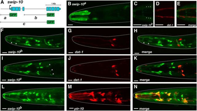
Expression pattern of swip-10. A, Summary diagram of constructs used for swip-10 promoter GFP fusion experiments. Data derive from L4 animals. PCR products were generated via overlap PCR as described in the Materials and Methods. B, GFP expression under the control of the swip-10a promoter in hypodermal cells. C–E, GFP expression driven by swip-10b is visible along processes (arrowheads) that run parallel to DA neuron dendrites as revealed by coexpression with dat-1: mCherry. F–H, swip-10b:GFP is expressed in a number of cells located in the head that do not overlap with DA neurons. Arrowheads in F denote processes similar to C–E. I–K, swip-10c:GFP is expressed in multiple head cells, include low expression in DA neurons (arrowheads). L–N, swip-10b:GFP expression (L) is colocalized in multiple cells with reporter driven by the pan-glial promoter ptr-10 (M). Scale bars, 10 μm in all images. Dotted lines denote the outline of the worm head as revealed by DIC imaging.

