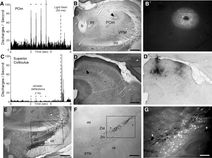
Figure 2.
Light-responsive region in superior colliculus projects to ZIv region that innervates whisker-responsive region in POm. A, PSTH shows whisker-induced multiunit responses recorded in POm by an FG-filled pipette. This site did not respond to a 50 ms light flashed in both eyes. B, Section through POm indicates necrosis at the FG deposit marking the whisker-responsive site in A. B′, Same section under fluorescent illumination shows extent of the FG deposit. C, Multiunit responses to visual light flash recorded in the medial superior colliculus by a BDA-filled pipette. Neurons at this site responded weakly to whisker stimulation. D, CO-processed section of the recording site (arrow) in the medial superior colliculus. D′, Adjacent section shows the BDA deposit marking the light-responsive site in C. E, CO-processed section through ZI and STN. Inset indicates region displayed in F. F, Adjacent section shows BDA-filled cells and terminals in upper part of ZIv. Inset indicates the region displayed in G. G, Fluorescent illumination at high-magnification reveals BDA-labeled terminals intermingled with FG-labeled neurons in ZIv. Arrows indicate same blood vessels in F and G. Scale bars: B, B′, D, D′, E, 500 μm; F, 250 μm; G, 100 μm.

