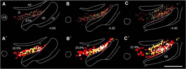
Figure 4.
Collicular projections overlap the ZI neurons that project to POm. A–C, Reconstructed plots of FG-filled neurons (gold) and FR-labeled terminal varicosities (red) in ZI. Numbers indicate distance from bregma in millimeters. A′–C′, Overlap analysis of FG-labeled neurons and FR-labeled terminals in ZI, using 50 μm2 bins. Each panel corresponds to the plotted reconstruction shown directly above. For the overlap analysis, gold bins contain at least one FG-labeled neuron, red bins contain at least two FR-labeled terminals, and white bins contain at least one FG-labeled neuron and two FR-labeled terminals. Percentages indicate proportion of white bins. Scale bar, 1 mm. cp, Cerebral peduncle; ml, medial lemniscus; mt, mammilothalamic tract; ot, optic tract.

