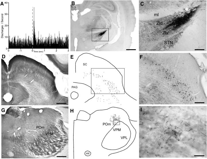
Figure 6.
Afferent and efferent connectivity of a visually responsive region in ZIv. A, PSTH shows neural responses in lateral ZIv to a 50 ms visual light flash recorded by a pipette filled with both FG and BDA. B, C, BDA deposit at the light-sensitive site recorded in A. D, CO-processed section through the superior colliculus. E, F, Plotted reconstruction and photomicrograph of BDA-labeled neurons in superior colliculus; rectangle indicates region in F. G, CO-processed section through POm and VPM. H, I, Plotted reconstruction and photomicrograph of BDA-labeled terminal varicosities in POm; rectangle indicates the region in I. Scale bars: B, D, G, 500 μm; F, 250 μm; C, 100 μm; I, 50 μm. PAG, Periaqueductal gray; VPM, ventroposteromedial; VPL, ventroposterolateral.

