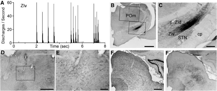
Figure 7.
Afferent and efferent connections of a whisker-responsive region in ZIv. A, PSTH shows neural responses in ZIv recorded by a FG/BDA-filled pipette during whisker deflections administered at 2, 5, and 8 Hz. B, C, BDA deposit at the site of the whisker-responses recorded in A. Rectangles in B indicate regions in C and D. D, E, Low- and high-power views of BDA-labeled axonal terminals in POm. D, Inset indicates region in E. F, Thionin-stained section through the superior colliculus. F′, Adjacent section showing the laminar location and spatial extent of BDA-labeled neurons in the superior colliculus. Scale bars: B, 1 mm; D, F, 500 μm; C, 250 μm; E, 100 μm.

