
Figure 9.
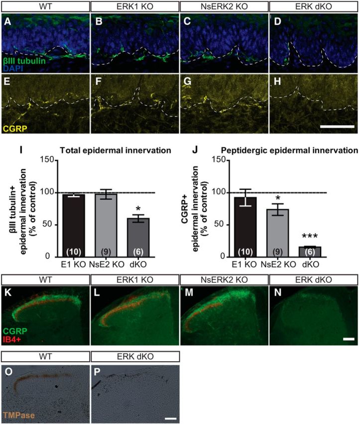
Expression of at least one ERK isoform is necessary for target innervation of epidermis and SCDH. A–D, Representative confocal images of total IENFD demonstrate βIII-tubulin+ fibers coursing through dermal–epidermal border in WT (A), ERK1 KO (B), NsERK2 KO (C), and ERK dKO (D) mice. E–H, Representative confocal images of peptidergic IENFD demonstrate CGRP+ fibers coursing through dermal–epidermal border in WT (E), ERK1 KO (F), NsERK2 KO (G), and ERK dKO (H) mice. Scale bar, 50 μm. I, J, In independent studies, average total (I) and peptidergic (J) IENFD (fibers/100 μm) were determined for NsERK2 KO (n = 9), ERK1 KO (n = 10), and ERK dKO (n = 6) mice, and then data were divided by the control (n = 6–10) littermates' IENFD for graphical illustration (mean ± SEM). K–N, Representative images of nociceptive central termination in the SCDH demonstrated CGRP+ (green) and IB4+ (red) nociceptive terminals in laminas I and II, respectively, for WT (K), ERK1 KO (L), NsERK2 KO (M), and ERK dKO (N) mice. Scale bar, 100 μm. TMPase assay assessed PAP activity present in nonpeptidergic fibers terminating in lamina II of SCDH in WT (O; n = 4). P, ERK dKO (n = 4) did not exhibit any TMPase activity indicative of nonpeptidergic terminals. Scale bar, 100 μm. *p < 0.05 and ***p < 0.001.