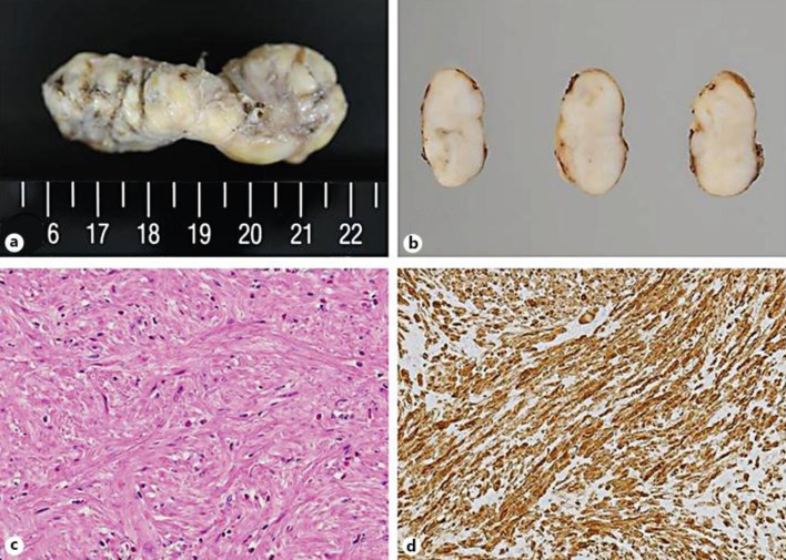
Fig. 5.
Pathological features of the tumor. a Grossly, the tumor had an elongated dimension, presenting as a sausage-shaped mass. b The cut surfaces were of a grayish tan. c Microscopically, the tumor was composed of well-differentiated smooth muscle cells. Neither mitotic figures nor necrotic foci were detected in the tumor. HE. ×200. d Immunohistochemically, the tumor cells were positive for desmin. They were almost negative for KIT.

