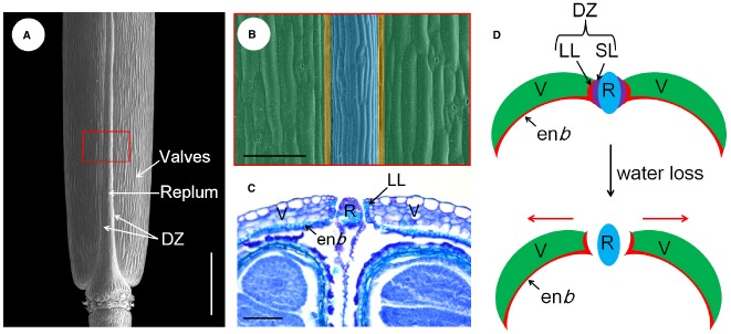
FIGURE 1.

Tissue organization and pod dehiscence process of the Arabidopsis fruit. (A) Scanning electron microscopic (SEM) micrograph of a mature silique, the different parts are indicated. (B) A close-up view of the red boxed area shown in (A), the valve, DZ, and replum are shaded with green, yellow, and blue color, respectively. (C) transversal section of the ovary region of a mature silique showing the SL has already been disintegrated and the silique opens from the replum. (D) Models for the pod dehiscence process of Arabidopsis, not to scale. The red arrows indicate the mechanical force generated in the valves. DZ, dehiscence zone; enb, endocarp b layer; LL, lignified layer; R, replum; SL, separation layer; V, valves. Scale bars in (A), 1.5 mm; (B,C), 80 μm.
