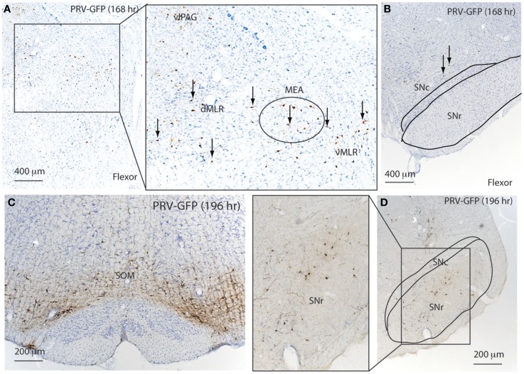
Figure 6.
Temporal and spatial distribution of PRV-GFP from the tibialis anterior (TA; flexor) muscle. At 168 h after PRV-GFP injections in TA, PRV-GFP preferentially labels the PAG, MLR, and MEA (A) as well as the VMM, but not the SNr (B). After 196 h, the PRV labeling from the TA shows more extensive labeling in the VMM (C) and SNr (D).

