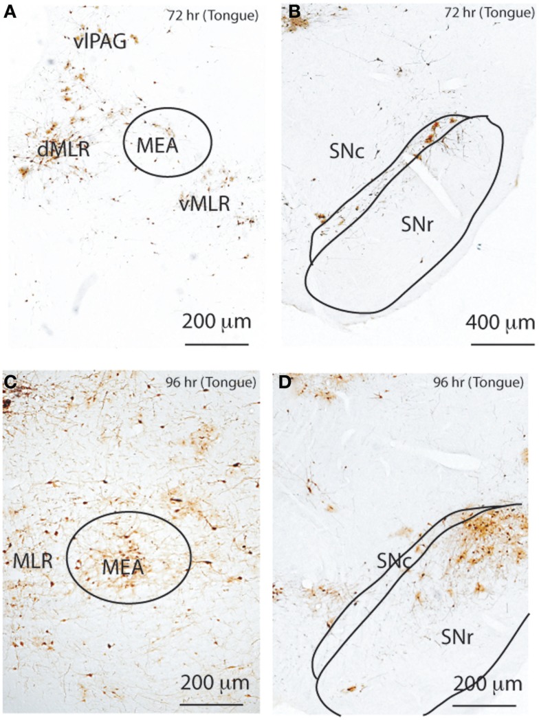
Figure 8.
Temporal and spatial distribution of PRV-GFP from the tongue. At 72 h after PRV-GFP injection, the MLR (A) and SNc (B) show PRV-GFP-ir neurons ipsilateral to the injection site. After 96 h, the labeling is more extensive (C,D), now including the MEA (C) and SNr (D). The temporal sequence of PRV labeling suggests that the SNc- > MLR pathway requires less time for labeling than the SNr- > MEA- > MLR pathway following tongue injections.

