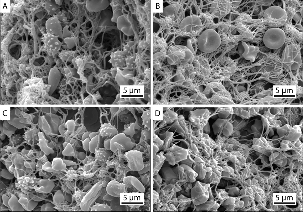
Figure 3.
Representative scanning electron microscopy images of (A, C) bare platinum coil–clot complexes and (B, D) clots alone. All images are at 3500× magnification. Echinocytes (speculated red blood cells) are present due to the diminishing supply of ATP during blood clot formation and storage at 5°C.

