
Figure 4.
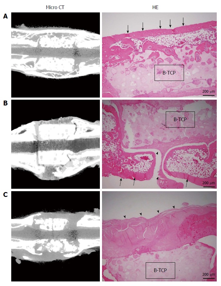
Micro-computed tomography images and hematoxylin and eosin-stained sections at 8 wk post-implantation. A: BMSC/TCP/Sheet constructs with continuous bone formation to host bone. Both micro-computed tomography (CT) and histology showed bridging bone formation over the TCP to host bone (arrows); B: BMSC/TCP/Sheet constructs with segmental bone formation over the TCP. Soft tissue interposition was observed between the TCP and the callus (arrowheads); C: BMSC/TCP constructs implanted into femurs. The TCP cylinders of the BMSC/TCP constructs were broken. Arrowheads indicate soft tissue interposition between the TCP and the autologous femur. BMSCs: Bone marrow stromal cells; TCP: Tricalcium phosphate.