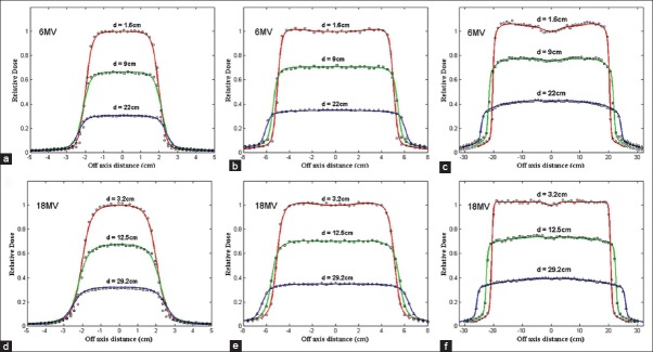
Figure 3.

Comparison of lateral dose distribution in a water phantom measured using a semiflex PTW ion chamber (shown in curves) and Monte Carlo calculated (shown in symbols) at 1.6, 9, and 22 cm depths in water for 6-MV photon beams and at 3.2, 12.5, and 29.2 cm depths in water for 18-MV photon beams. For both energy SSD was 100cm. (a) 6-MV and 4 × 4 cm2 field size, (b) 6-MV and 10×10 cm2 field size, (c) 6-MV and 40 × 40 cm2 field size, (d) 18-MV and 4 × 4 cm2 field size, (e) 18-MV and 10 × 10 cm2 field size, and (f) 18-MV and 40 × 40 cm2 field size
