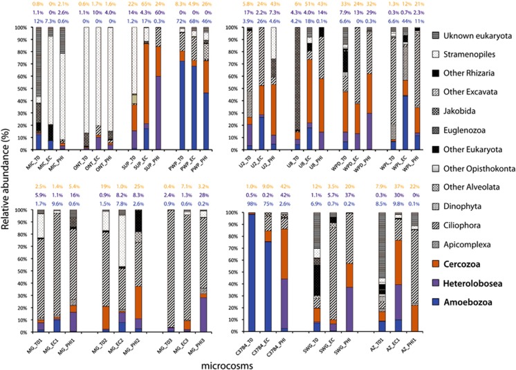
Figure 1.
Impact of L. pneumophila on the structure of eukaryotic microbial communities in laboratory microcosms. (a): Taxonomic affiliation of protist OTUs retrieved from different environmental samples incubated in the presence of E. coli (EC) or L. pneumophila str. Philadelphia-1 (PHI). Bars represent the relative abundance of protist taxa after rarefaction to correct for uneven sampling as described in the Materials and methods section. Sample identities are given on the x axis. MIC: fresh-water sample from Lake Michigan. ONT: fresh-water sample from Lake Ontario. SUP: fresh-water sample from Lake Superior. PWP: Pond-Washington Park (Chicago, IL, USA). U2: soil from Aguascalientes (Mexico). U8: soil from Neyaldi (India). WPD and WPR: soil samples from Washington Park (Chicago, IL, USA). MG: soil from Hyde Park (Chicago, IL, USA). C37B4: Core sample from ocean subsurface, Expedition IODP 311. SWG: Sewage (WWTP, Chicago, USA). AZ: soil from Tucson, AZ, USA. Sample-name_0: microcosm analyzed before the addition of E. coli or L. pneumophila. Sample-name_EC: microcosm incubated with 107 cells of E. coli. Sample-name_PHI: microcosm incubated with 107 cells of L. pneumophila Philadelphia 1. Relative abundance values (%) of Cercozoa, Heterolobosea and Amoebozoa are highlighted in orange, purple and blue color, respectively. See Microcosm design in Materials and methods Section for details.

