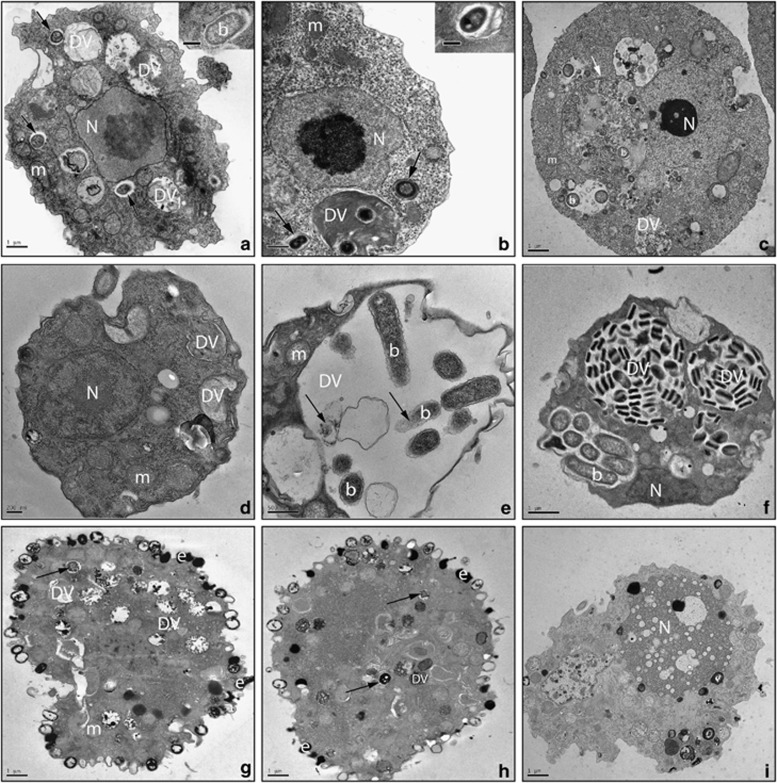
Figure 3.
Protist grazers on L. pneumophila. Transmission electron micrographs of isolates S. palustris (a–c), Paracercomonas CWPL (d–f) and Cercomonas MG33 (g–i) showing control preparations with ingested E. coli (a, d, g) and those fed with L. pneumophila JR32 (b, e, h) or L. steelei IMVS3376 (c, f, i). TEM micrographs were taken 48 h after the addition of Legionella to the protist. b: bacteria; DV: digestive vacuole; e: extrusomes; m: mitochondria; N: nucleus. Black arrow indicates bacterial cell being digested. White arrow highlights an autophagosome. See Supplementary Figure S5 for more evidence of cellular damage in S. palustris trophozoites grazing on L. steelei.

