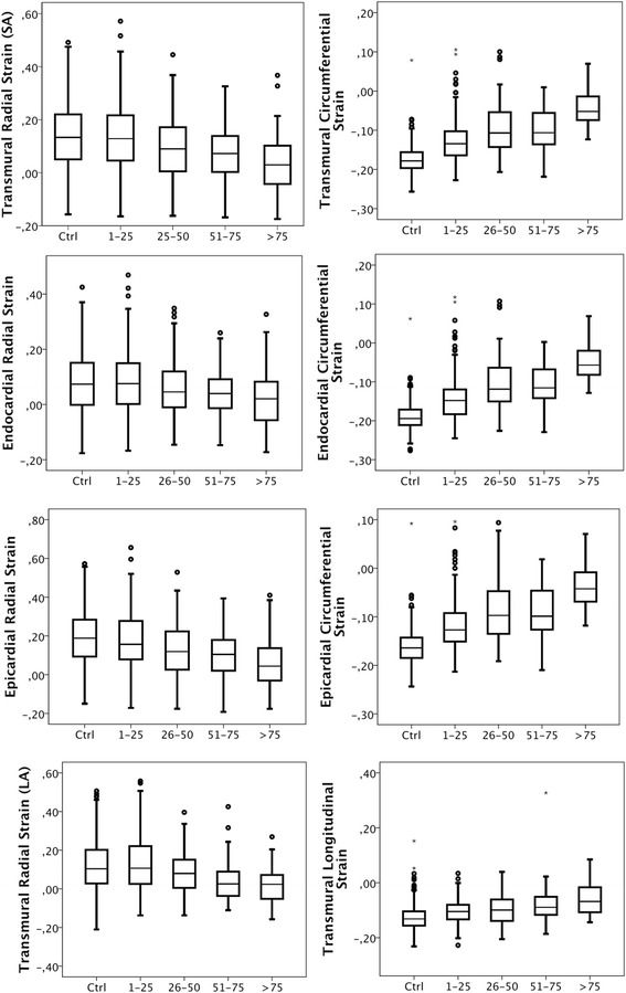
Fig. 1.

Layer strain versus segmental transmurality of scar. The box plots show median, the two central quartiles in the box, one quartile in each wisker and outliers. The upper three rows show radial (left) and circumferential (right) strain boxplots obtained from transmural (top), subendocardial (2nd from top) and epicardial (3rd from top) measurements. The fourth row shows transmural radial (left) and lonitudinal (right) strain obtained from the long axis in segments with various degree of transmurality of scar
