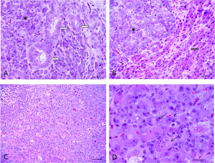
Fig. 1.
Histologic section from the specimen from the liver of the dog. HE stain (A, B). Hepatocellular carcinoma (*) and cholangiocellular carcinoma (arrow) (A). In part, hepatic cells with a normal pattern were observed (arrow). The tumor cells contained various-sized cord-like structures and conspicuous nucleoli (*). Bar=20 µm. (B). Histologic section of the nude mouse with a transplanted tumor. HE stain (C, D). Bar=100 µm (C). The cells of 2 nuclei were observed in various-sized and inequality (arrow). Bar=20 µm (D).

