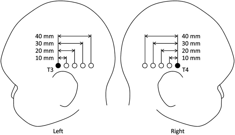
Fig. 1.
Arrangement of the light sources and detectors in near-infrared spectroscopy (NIRS) measurements for hemodynamic responses to auditory stimuli in infants. Filled and open circles show the positions of the light sources and detectors, respectively. The source optical fibers were placed on the T3 and T4 positions of the International 10/20 system. The position of the measurement channel for recording the changes in oxygenated and deoxygenated hemoglobin (oxy- and deoxy-Hb) signals is defined as the midpoint between the source and detector. Four different detectors placed in a line on each hemisphere detect reflected light from a single incident position. Thus, hemoglobin signals are simultaneously measured by a total of eight channels. The data obtained at source-detector 40-mm channel were not used in the analysis.

