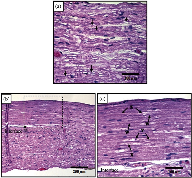
Fig. 3.
Histological comparison of safe versus nonsafe of optically stimulated experimental sites in dorsal lumbar nerve roots. (a) Experimental site ( magnification) resulting in the stimulated muscle recordings using for a total of 20 laser pulses (). Numerous axons (arrows) can be seen in the nerve fibers in this image. (b) An overview of the thermal lesion produced within the dorsal root nerve ( magnification) using (). The lesion is generally hyperchromatic and the endoneurial tubes are straightened out in the center. The arrow represents the interface between the thermally damaged tissue and the underlying normal tissue. (c) The demarcated area from (b) at magnification. This lesion has the following features characteristic of thermal damage: swelling and hyperchromasia of the collagen in the endoneurium (W), granular degeneration of myelin (X), straightening of the nerve fibers as compared to the intact fibers at the bottom of the image, vacuolization, and expansion of the endoneurial tubes in some areas (X). Although some axons show axonal thermal damage (Y), others in the lesion (Z) do not at this level of magnification.

