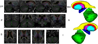
Fig. 3.

Overlay of surface outlines on individual slices of T1 MRI in the three orthogonal views along with the surface representation for a representative subject in the template library. [Cerebellum (green), thalamus (magenta), caudate (turquoise), hippocampus (blue), putamen (red), and lateral ventricles (yellow).]
