
Fig. 12.
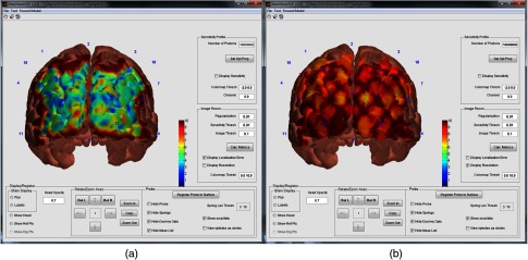
(a) The localization error map for the square probe geometry from Sec. 3.1. (b) The resolution map for the same probe. Variation in localization error is highly dependent on the underlying anatomical structure, while probe resolution is predominately a function of source and detector placement. The color scale is linear and spans from 0 to 10 mm as indicated in the “Colormap Thresh” edit box.