
Fig. 7.
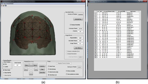
(a) The AtlasViewer graphical user interface provides a number of tools for viewing probe geometries: the cortical surface may be colorized using a variety of color maps to depict different anatomical regions, the head surface may have its opacity adjusted or removed entirely, 10-20 reference points may be toggled on/off, the head may be rotated and zoomed, and various elements of the SD file may be hidden or revealed. (b) The popup window that is created when the “Project to cortex” button is pressed contains the list of SD pairs in the measurement list, the channel coordinates in the Monte Carlo space, the channel coordinates in the Montreal Neurological Institute (MNI) space, and a label for the cortical surface that these MNI coordinates correspond to in the segmented atlas volume. Channel coordinates are given at the midpoint between the associated source and detector.