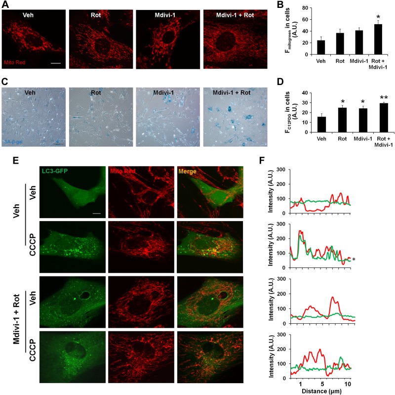
Figure 4.
Accumulation of damaged mitochondria leads to cellular senescence in lung fibroblasts. A) Representative images of mouse lung fibroblasts show mitochondrial elongation and accumulation of mitochondrial mass with rotenone (Rot) and Mdivi-1 treatment. Rotenone (10 nM), Mdivi-1 (1 µM), and their combination for 15 days with alternate day treatment are shown. Veh, vehicle. B) Average fluorescent intensity of MitoTracker Green (Fmitogreen) reflects mitochondrial mass, measured by FACS in mouse lung fibroblasts. A.U., arbitrary units. *P < 0.05 vs. vehicle. C) Representative images of SA-β-gal activity in mouse lung fibroblasts with and without rotenone (10 nM), Mdivi-1 (1 µM), and combination treatment. D) C12FDG fluorescence in mouse lung fibroblasts was measured by FACS and plotted as average fluorescent intensity (FC12FDG in cells), which showed the degree of cellular senescence in arbitrary units. *P < 0.05 and **P < 0.01 vs. vehicle. E) Representative images of mouse lung fibroblasts show colocalization of LC3-GFP with mitochondria (Mito Red). Vehicle or rotenone- and Mdivi-1-treated cells were further treated with or without CCCP for 2 hours before imaging. F) Line scan of fluorescence intensity in the corresponding images. *P < 0.05 vs. control. Data are shown as the means ± sem (n = 3–4). Scale bars, 10 μm (A and E) and 100 μm (C).

