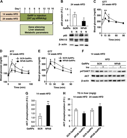
Figure 5.
GeRP-mediated silencing of Nf-κb in KCs in mice fed a HFD improves glucose tolerance. A) Protocol of GeRP treatment in mice fed an HFD for 14 or 24 weeks. B) Representative Western blot and protein levels of NF-κB normalized to β-actin levels in KCs following a 15-day GeRP treatment in mice fed 24 weeks of an HFD. ERK1/2 was used as a negative control (S, SCR; N, NF-κB; n = 5). C) GTT (1 g/kg) was performed in mice fed 24 weeks of an HFD and treated with SCR- or NF-κB-GeRPs (n = 11–14). D) PTT (1 g/kg) was performed in mice fed a 24-week HFD treated with SCR- or NF-κB-GeRPs after withholding food for 6 hours (n = 5). E) GTT (1 g/kg) was performed in mice fed a 14-week HFD and treated with SCR- or NF-κB-GeRPs (n = 11–14). F) Representative Western blot of total and activated (pSer473) Akt in the liver of 14-week HFD mice. G) F.C. of Akt phosphorylation by insulin measured by densitometry of pSer473-Akt normalized to total Akt (n = 9–10). H) TG content in the liver (n = 5). Results are presented as mean of F.C. normalized to SCR-GeRPs–treated mice ± sem. *P < 0.05; **P < 0.01; ***P < 0.001. The statistical significance was analyzed by t test or ANOVA followed by Tukey posttest.

