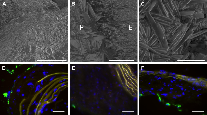
Figure 2.
A) Scanning electron microscopy of cholesterol crystals densely deposited on surface of aortic plaque. Scale bar, 100 μm. (B) Scanning electron microscopy of cholesterol crystals on denuded plaque (P) but not on adjacent regular endothelium (E). Scale bar, 40 μm. C) Higher magnification scanning electron microscopy depicts morphology of cholesterol crystals. Scale bar, 20 μm. D–F) Immunofluorescent staining for CD31 (green) demonstrates little to no intraplaque angiogenesis in ApoE null fed a Western diet for (D) 4 months, (E) 5 months, and (F) 6 months. Scale bar, 50 μm.

