
Figure 1.
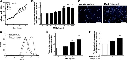
TRAIL stimulates human preadipocyte proliferation. A–C) SGBS preadipocytes were stimulated with growth medium alone or with increasing doses of TRAIL. A) At the indicated time points, the number of adherent cells was counted using a net micrometer. Data are expressed as means ± sem of 3 independent experiments performed. *P ≤ 0.05 for TRAIL 100 ng/ml compared with growth medium alone at 72 hours. B) After 72 hours, [3H]-thymidine incorporation was measured. Data are expressed as means ± sem of at least 3 independent experiments performed. **P ≤ 0.01; ***P ≤ 0.001 compared with growth medium alone. C) After 72 hours, Hoechst 33342 fluorescence staining was performed. One representative of 3 independent experiments performed is shown. Scale bars, 200 µm. D) SGBS preadipocytes were stained with CFSE and stimulated with basal medium, growth medium alone or with TRAIL. After 72 hours, the remaining CFSE fluorescence was measured. A representative histogram of 1 of 3 independent experiments performed is shown. The loss of CFSE mean fluorescence intensity in comparison to cells stimulated with basal medium was calculated. *P ≤ 0.05 for TRAIL 30 ng/ml compared with growth medium alone. E) Human primary stromal-vascular cells were isolated from adipose tissue samples of 4 patients and stimulated with growth medium alone or with increasing doses of TRAIL. After 72 hours, [3H]-thymidine incorporation was measured. Data are expressed as means ± sem. *P ≤ 0.05; **P ≤ 0.01 compared with growth medium alone. F) SGBS preadipocytes were stimulated with growth medium alone or human agonistic antibodies specific for either TRAIL-R1 (mapatumumab, mapa) or TRAIL-R2 (lexatumumab, lexa). After 72 hours, [3H]-thymidine incorporation was measured. Data are expressed as means ± sem of 3 independent experiments performed.