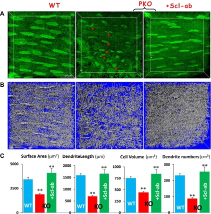
Figure 4.
Statistically analyzing morphologic changes of Ocys in PKO jawbones with and without Scl-Ab treatment. A) Confocal FITC image shows spindle-shaped Ocys with numerous dendrites in a well-organized arrangement in the WT (left) compared to the PKO Ocys with vehicle (middle) or Scl-Ab treatment (right). B) Artificial silver images reconstructed with the Imaris software from (A) are shown. C) The statistical analysis using Imaris software reveals significant differences in Ocy surface area, dendrite length, total cell volume, and dendrite numbers among WT, PKO, and PKO Ocys treated with Scl-Ab for 8 weeks. **P < 0.01 (n = 20).

