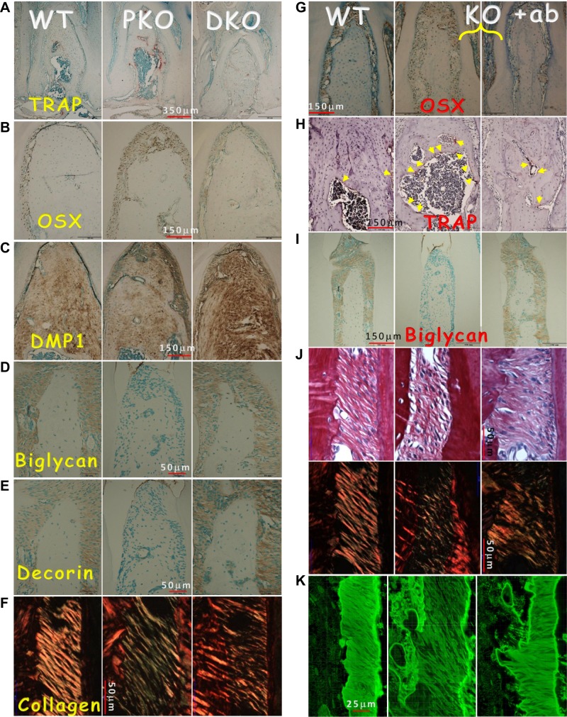
Figure 6.
Restoration of the molecular markers and collagen in the DKO mice and the PKO mice treated with Scl-Ab for 8 weeks. A) TRAP stains reveal a reduction of osteoclast number in DKO jawbones. B–E) Recovery of molecular markers in DKO mice by immunostaining, including OSX (B), DMP1 (C), biglycan (D), and decorin (E), as well as partial restoration of collagen (F) in DKO using polarized light under the microscope. G) Representative OSX immunostains in WT (left), PKO (middle), and PKO treated with Scl-Ab (right) are shown. H) TRAP stain images in 3 groups are shown. Yellow arrow indicates osteoclast (stained red by TRAP). I) Biglycan stain images in 3 groups are shown. J) Sirius stain images by regular microscopy (upper) and polarized microscopy (lower) in the 3 groups are shown. K) FITC stain images in the 3 groups are shown. The data show improvement in all these markers and PDL fibers in the Scl-Ab-treated group.

