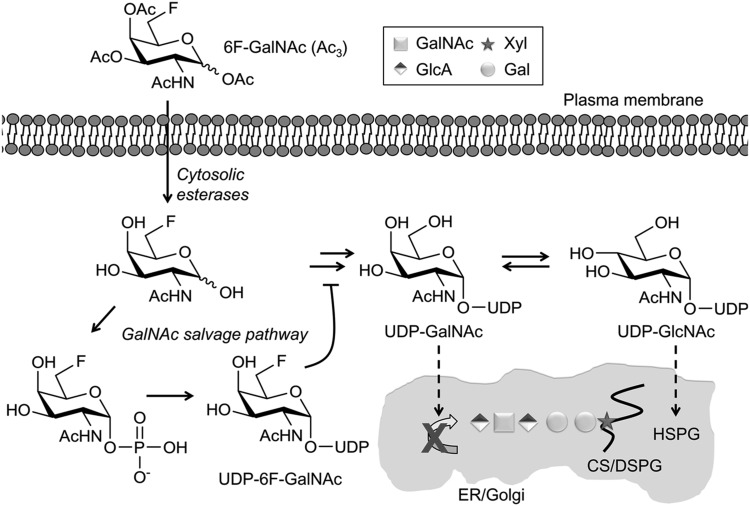
Figure 6.
A sugar-nucleotide-mediated mechanism of inhibition of GAG biosynthesis by 6F-GalNAc (Ac3). Proposed mechanism: 6F-GalNAc (Ac3) enters the cell by passive diffusion, is deacetylated by aspecific cytosolic esterases, and becomes activated into UDP-6F-GalNAc by the machinery of the GalNAc salvage pathway. 6F-GalNAc is not incorporated into GAGs, but reduces the pool of natural UDP-GalNAc by using the same activation machinery or by a feedback mechanism. UDP-GlcNAc is also reduced, because UDP-GalNAc and UDP-GlcNAc are in equilibrium, catalyzed by an epimerase. It is likely that a part of UDP-6F-GalNAc is converted into UDP-6F-GlcNAc via the same epimerase reaction, thereby possibly contributing to the reduced level of UDP-GlcNAc via a nucleotide sugar feedback mechanism (18). As a result, the biosynthesis of GAGs and other glycans is reduced. The curving line represents a core protein. DSPG, chondroitin sulfate/dermatan sulfate proteoglycan. HSPG, heparan sulfate proteoglycan.

