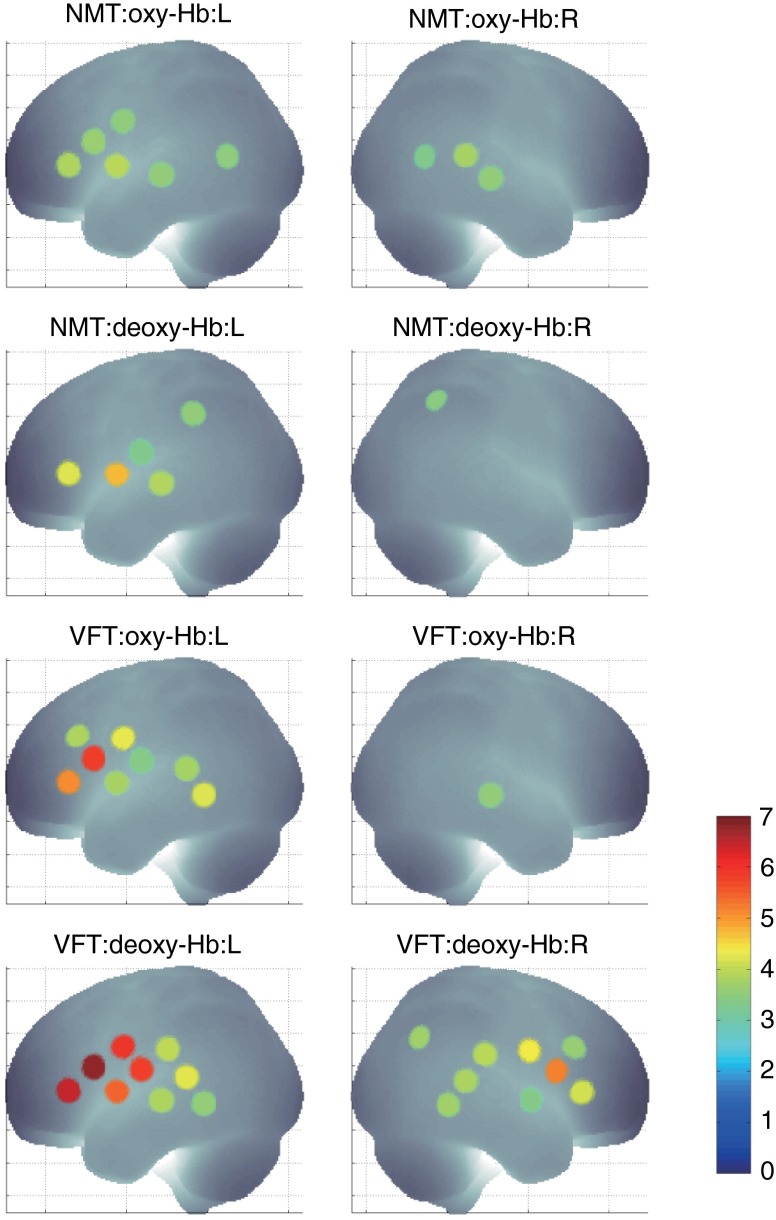
Fig. 5.
The results of the group analysis using the optimized value. The channels with significant -values are presented as described for Fig. 4 (, Bonferroni corrected). From top to bottom, the maps respectively indicate analyses of the oxy-Hb signal for the NMT, the deoxy-Hb signal for the NMT, the oxy-Hb signal for the VFT, and the deoxy-Hb signal for the VFT. Significant activations were detected for all conditions on both hemispheres.

