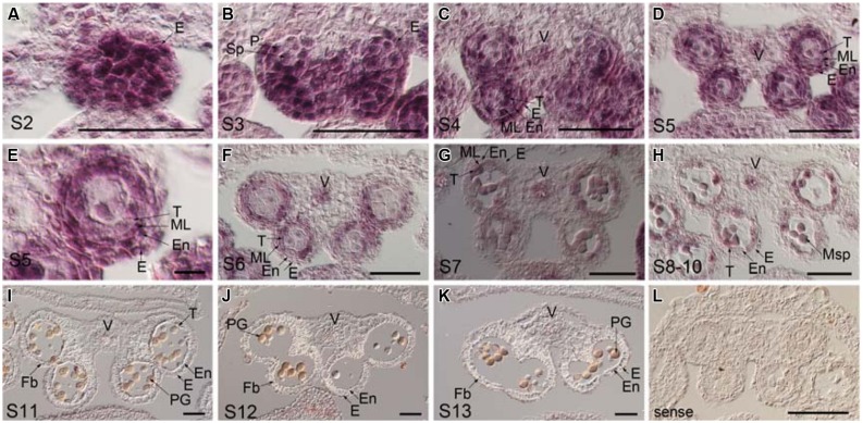
FIGURE 5.
Temporal and spatial expression patterns of TCP24 gene during anther development. In situ hybridization using TCP24 probe. (A–K) Transverse sections of anthers at the stages 2 (A), 3 (B), 4 (C), 5 (D,E), and 6 (F), 7 (G), 8–10 (H), 11 (I), 12 (J), and 13 (K) antisense probes. (E) Magnified picture from the top right region in (D). (L) Sense probe of TCP24. E, epidermis; En, endothecium; Fb, fibrous bands; ML, middle layer; Msp, microspore; P, parietal cell; PG, pollen grain; Sp, sporogenous; T, tapetum; V, vascular region. Scale bars: 50 μm in (A–D, F–L); 5 μm in (E).

