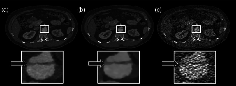
Fig. 3.
62-year-old male patient with an aortic dissection. The panels (A–C) from left to right: , IMBR, and FBP reconstruction. The effective dose was 2.32 mSv. The strongly eliminated noise enables the detection of the aortic dissection in panels A and B, while being hard to detect in panel C. (window width, 1200 HU; window center, 200 HU).

