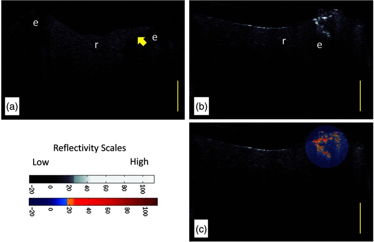
Fig. 2.
(a) CP-OCT grayscale intensity image of a resin composite filling (r) placed recently within 2 to 6 months (Subject 823). The enamel (e) below the margin of the restoration shows low-backscattered intensity. (b) CP-OCT grayscale intensity image of an interface with cavitated secondary caries. (c) A second false color scale CP-OCT image is overlaid on the grayscale image to identify areas where the enamel scattering is over 13.3 dB (orange shades) and 30.1 dB (red shades). Yellow scale bar is 1 mm of optical depth.

