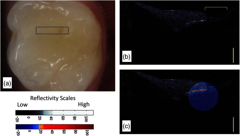
Fig. 5.
(a) The margin of this recently placed restoration on tooth A (subject 820) had a marginal defect area with marginal discoloration. (b) The CP-OCT image a definitive area (brackets) where the restoration is not well adapted to the tooth, possibly from postplacement fracture. (c) A second false color scale CP-OCT image is overlaid on the grayscale image to identify areas where the enamel scattering is the enamel scattering is over 17.5 dB (orange shades) and 25.9 dB (red shades). Yellow scale bar is 1 mm of optical depth.

