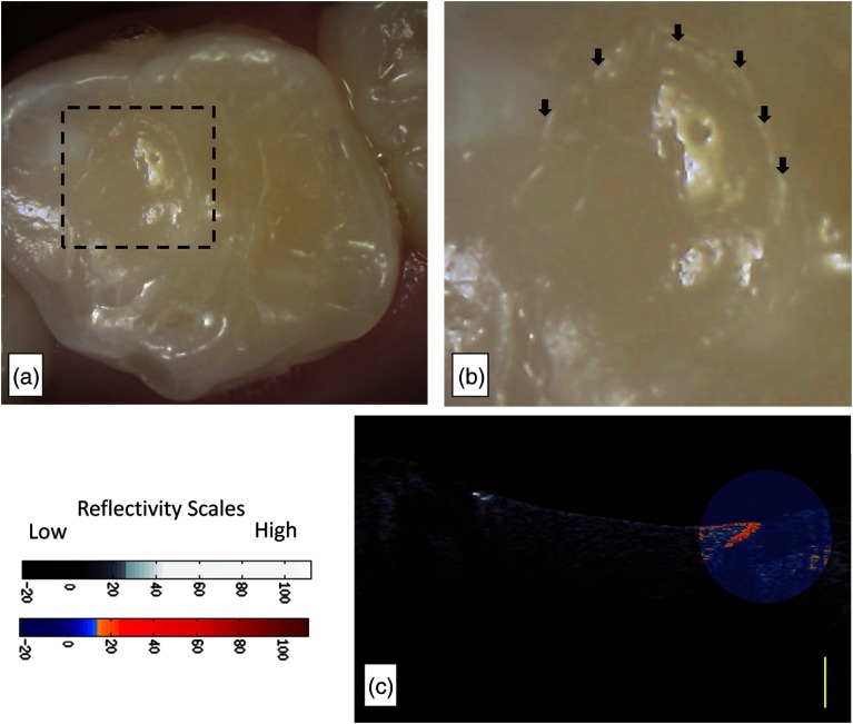
Fig. 6.
Subject 847 had a restoration where the subsurface backscattering was 20.03 dB. (a) Intraoral image of a resin composite restoration on the occlusal of Tooth J placed 61 days from the day of assessment. (b) The visual examination noted marginal opacity which can be seen in the intraoral image. There is a pervasive marginal opacity around the margins of the restoration (arrows). (c) The CP-OCT images revealed a subsurface region of increased scattering and depolarization that was graphed. A second false color scale CP-OCT image is overlaid on the grayscale image to identify areas where the enamel scattering is the enamel scattering is over 13.3 dB (orange shades) and 23.8 dB (red shades). Yellow scale bar is 1 mm of optical depth.

