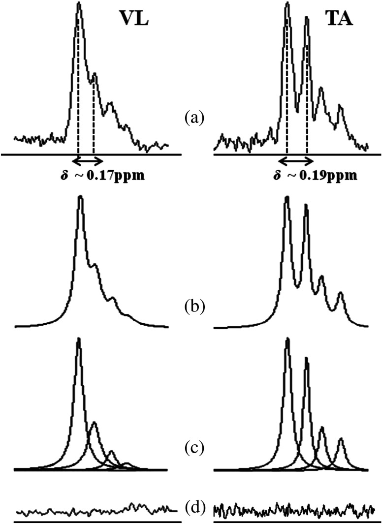
Fig. 4.
spectrum of the VL and TA muscles processed by AMARES. (a) Chemical shift () estimated from diffusion tensor imaging (DTI) measurements of these muscles were used in prior knowledge constraints for spectral fitting. (b) Fitted estimate spectrum, (c) individual components, and (d) residues can also be seen. Better separation between IMCL and EMCL peaks were seen between TA and were well-agreed with derived from DTI.

