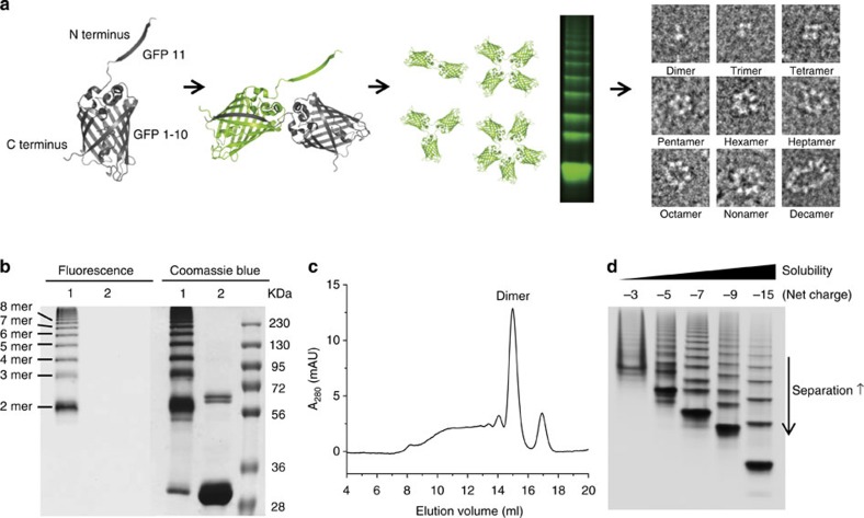
Figure 1. Cellular self-assembly of GFP oligomers.
(a) Schematic representation of the fabrication of discrete GFP (nano)polygons. The β strand 11 of GFP is connected to the N terminus of GFP 1–10 using a short peptide linker, and the resulting GFP monomer unit undergoes self-assembly into polygonal structures in the cell. Through the introduction of supercharges on the polygon surface, GFP polygon mixtures can be separated and isolated depending on the number of GFP-building blocks, to produce a series of discrete GFP polygons. (b) SDS–PAGE analysis of GFP oligomers. Oligomer mixtures were applied to a PAGE gel containing 0.1% SDS without (lane 1) or with (lane 2) boiling. The gel was analysed by a fluorescent image analyser with 470-nm excitation and 530-nm emission filters (left), and Coomassie blue staining (right). (c) SEC of GFP oligomers using a Superdex 200 column (10/300 GL). (d) Native PAGE analysis of GFP oligomer charge variants with net charges of −3, −5, −7, −9 and −15. Enhanced solubility and gel separation of the oligomers are indicated.

