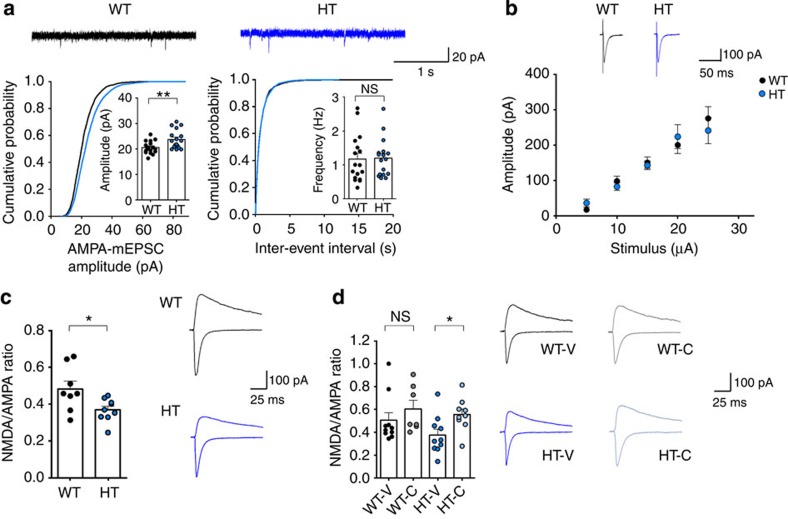
Figure 8. CQ restores NMDAR function at Tbr1+/− amygdalar thalamic-LA (T-LA) synapses.
(a) CQ increases mEPSC amplitude but not frequency in Tbr1+/− principal neurons in the LA (4–6 weeks). (n=17 cells (3 animals) for WT and HT, **P<0.01, Student's t-test). (b) CQ has no effect on the input–output ratio at Tbr1+/− T-LA synapses (4–6 weeks), as indicated by plots of fEPSP slopes against stimulus intensities. Representative current traces are an average of three consecutive responses with input stimulations of 25 μA. (n=8 cells (3 animals) for WT and HT; Student's t-test). (c) Reduced NMDA/AMPA ratio at Tbr1+/− T-LA synapses (4–6 weeks). (n=8 cells (5 animals) for WT and 9 (5) for HT; *P<0.05; Student's t-test). (d) CQ restores the NMDA/AMPA ratio at Tbr1+/− T-LA synapses but has no effect on WT synapses (4–6 weeks). (n=10 (5) for WT-V, 7 (4) for WT-C, 10 (6) for HT-V and 9 (4) for HT-C; *P<0.05; two-way analysis of variance (ANOVA) and one-way ANOVA with Tukey's post hoc test). Data in all panels with error bars represent mean±s.e.m. NS, not significant.

