
Fig. 1.
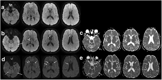
Brain MRIs of index case. a-e: MRI was normal at 4 months without clear evidence of restricted diffusion on DWI sequence (a), but progressed to restricted diffusion in the right caudate head (solid arrows) and more subtle cortical (dashed arrows) and possible superior vermis (dotted arrows) involvement at 10 months (b, DWI; c, ADC map), with progression to bilateral asymmetric caudate and more evident cerebellar vermis involvement at 11.5 months (d, DWI; e, ADC map)