
Fig. 3.
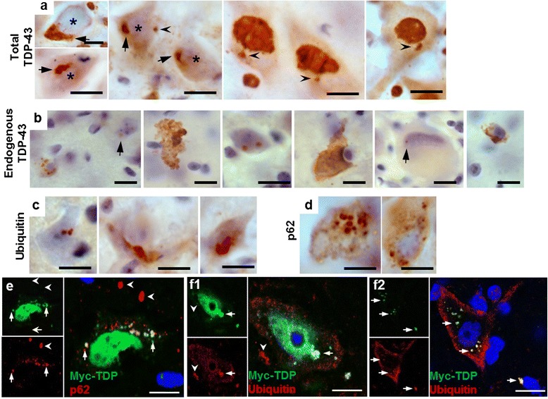
Co-expression of TDP-43WT and TDP-43Q331K in mice results in ubiquitin and p62 positive TDP-43 inclusions in the anterior horn of the lumbar spinal cord. a Large perinuclear TDP-43 aggregates (arrows) and small cytoplasmic TDP-43 inclusions (arrowheads) were present in multiple cells from the anterior horn of the lumbar spinal cord of TDP-43WTxQ331K animals. In many cases, these aggregates are accompanied by evidence of nuclear clearing of TDP-43 (asterisks) There is also evidence of large and small (arrows) cytoplasmic and nuclear inclusions of endogenous mouse TDP-43 (b), accompanied by ubiquitin (c) and p62 (d) aggregation in TDP-43WTzQ331K mice (scale bar: 10 μm). e–f Fluorescent co-labelling of myc-TDP and p62 (e) or ubiquitin (f) in the anterior horn of TDP-43WTxQ331K animals shows aggregates that are positive for both myc-TDP and p62/ubiquitin (arrows), together with myc-TDP negative, p62 or ubiquitin inclusions (arrowheads). TDP-43 and ubiquitin aggregates can be seen both in the presence (f1) and absence (f2) of nuclear TDP-43 (scale bar : 10 μm)