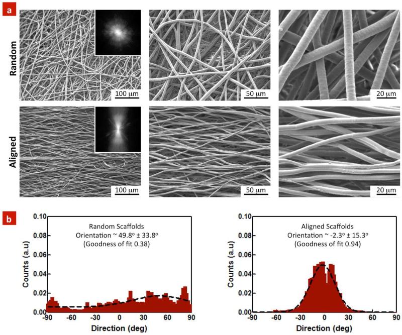
Figure 1. Topographical evaluation of random and aligned PGS-PCL scaffolds.
(a) SEMs images indicate arbitrary orientation of individual fibers within the random scaffolds, whereas highly organized and aligned fibers were observed in the aligned scaffolds. Inset: 2D FFT of SEM images further verified highly oriented structure within the aligned scaffolds. (b) The random scaffolds exhibited only a small degree of preferred orientation (49.8±33.8°), as compared to the aligned scaffolds (2.3±15.3°). The data represents fiber diameter calculated from at least 100 fibers (n=3).

