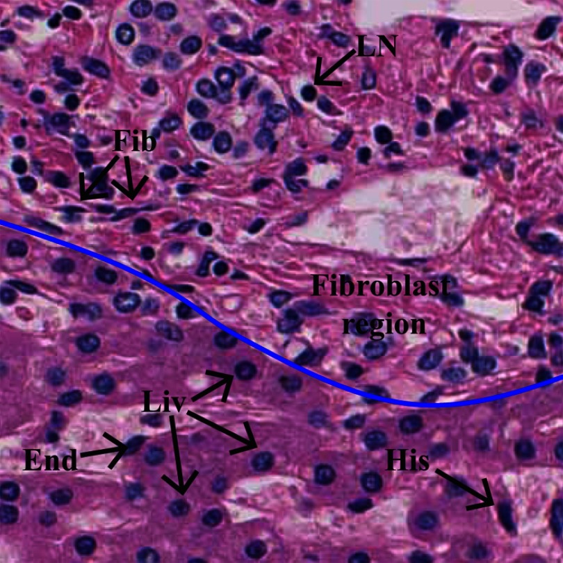
Fig. 2.
Description of HE-stained liver biopsy image. The image contains different types of cell nuclei and cellular components. Five types of nuclei have been annotated: Liv: liver cell nucleus, Fib: fibroblast cell nucleus, LN: lymphocyte, End: endothelium, and H: histiocyte. Upper part of the blue colored line indicates fibrous region.

