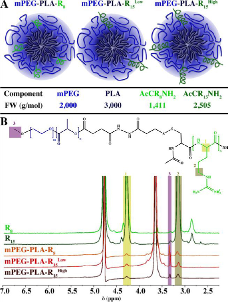
Figure 1. Micelles properties and surface oligoarginine presence.
(A) Schematic representation of the micelles formed and the relevant molecular weights of the components. (B) 1H NMR analysis of the peptides (CR8 and CR15) and micelles (mPEG-PLA-R8, mPEG-PLA-R15Low, and mPEG-PLA-R15High) showing the arginine α proton (1; δ = 4.40 ppm, 1H), δ protons (2; δ = 3.20 ppm, 2H), or ω-terminal methoxyl protons (arrow; δ = 3.40 ppm, 3H).

