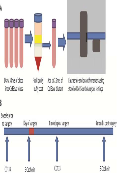
Figure A. Blood draw and experimental design.
(1) At each blood draw, blood was drawn into four CellSave tubes and within four hours, pooled into 30 mls whereupon the buffy coat, which contains CTCs, was purified by Ficoll gradient. The buffy coat was transferred to a new CellSave tube with CellSave dilution buffer to a total of 7.5mls, which was processed according to standard CellSearch procedures using the CTC enumeration kit. (2) The blood draw schedule is shown, including which marker was evaluated in the open channel of the CellSearch system at each draw.

