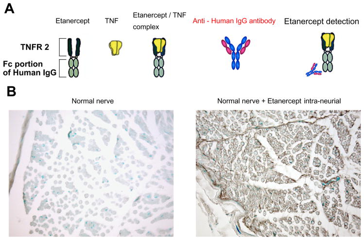
Fig. 1.
Establishment of the immunohistochemical method to detect the uptake and distribution of applied etanercept in rat nerves. (A) Model diagram of the detection technique for etanercept. (B) Immunohistochemical images of rat sciatic nerve stained with an antibody for human IgG. Methyl-Green nuclear counterstain was used. Normal nerve indicating relative lack of background antibody staining. Nerve 1 h following intra-neural injection of 125 μg of etanercept indicating its widespread distribution (brown). Representative micrographs of n=2 rats/group. Magnification 200×.

